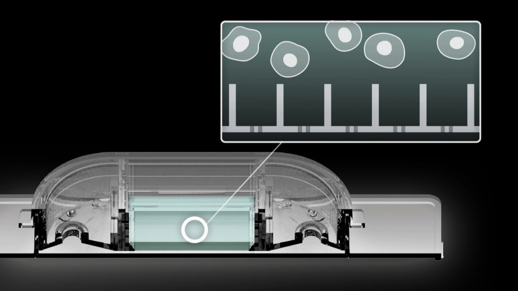
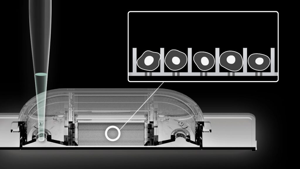
デバイス内での培養
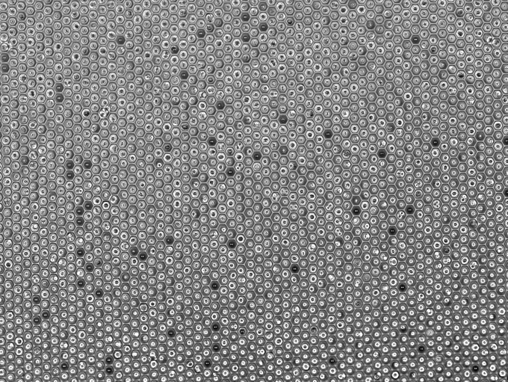
シングルセルとなるように分散した細胞を播種することで、ナノウェルの格納されたシングルセルの増殖等を観察可能です。
シングルセルとなるように分散した細胞を播種することで、ナノウェルの格納されたシングルセルの増殖等を観察可能です。
SIEVEWELL Slide is standard glass slide format and compatible with conventional microscope.

SIEVEWELL Slide は標準的なスライドガラスと同サイズです。 専用のアダプター等を必要とせず、一般的なチャンバースライドのようにそのまま顕微鏡でのイメージングが可能です。
HL-60 cells were cultured for 3 days with 1.3% DMSO to induce differentiation. Neutrophil-differentiated HL-60 cells were incubated for 5 minutes with the membrane-permeable NUCLEAR-ID Red DNA dye to stain nuclei. After three times of washing, NUCLEAR-ID Red stained-neutrophils were loaded into SIEVEWELL. RPMI containing Sytox Green (0.2 µM) and 100 ng/mL PMA was added to induce NETosis and to assess for cell death. SIEVEWELL was placed on IncuCyte® S3 System which is housed inside a cell incubator at 37°C with 5% CO2. Neutrophils were imaged using phase contrast, red (800 ms exposure) and green (400 ms exposure) channels. Images using a 20x dry objective lens were taken every 5 minutes.
Reference of reagents, scan conditions. J Immunol January 15, 2018, 200 (2) 869-879.
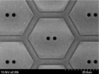
一つ一つのマイクロウェルの底部に約 2 μm の貫通孔が形成されています。中央のチャンバー部分に入れた細胞を含む溶液は、サイドポートからピペットで吸引することで吸い出すことができます。
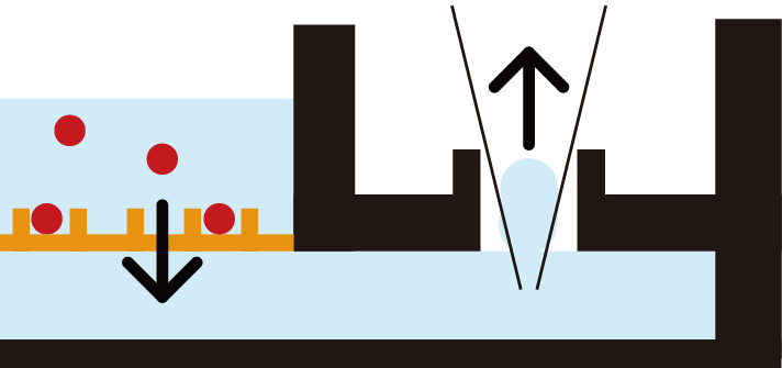
細胞は上から下に向かう溶液の流れに乗って、マイクロウェルに格納されます。
Step 1
細胞の懸濁液を中央のチャンバーに入れます
Step 2
サイドポートからバッファーを吸い出します
© Tokyo Ohka Kogyo Co., Ltd. 2024. All Rights Reserved